
🔍 Key Findings
Sample: 21 cats (10 control, 11 affected); 14 normal limbs vs 18 with MPL (MPL II: 7, MPL III: 11).
Significantly different CT measurements in MPL vs control:
- aLDFA: MPL II > control and MPL III (p = 0.014)
- FTW: MPL III > control (p = 0.021)
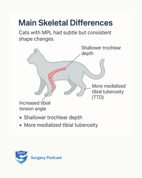
- FTD: control > MPL II and III (p < 0.001)
- TTA: MPL II and III had increased external tibial torsion vs control (p < 0.001)
- fPL and PV: MPL III cats had longer and more voluminous patellae
No significant differences in AA, mMPTA, TTD, fPW, aPH.
Patella width exceeded trochlear width in all groups.
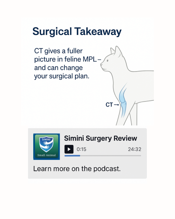
Authors suggest femoral and tibial angular correction may not be indicated in most feline MPL II–III cases.
Soft tissue techniques and trochleoplasty warrant further investigation.
CT method: Intraobserver ICC good in 64%, interobserver poor in 36% of metrics.


Simini Surgery Review Podcast
🔍 Key Findings
Sample: 21 cats (10 control, 11 affected); 14 normal limbs vs 18 with MPL (MPL II: 7, MPL III: 11).
Significantly different CT measurements in MPL vs control:
- aLDFA: MPL II > control and MPL III (p = 0.014)
- FTW: MPL III > control (p = 0.021)
- FTD: control > MPL II and III (p < 0.001)
- TTA: MPL II and III had increased external tibial torsion vs control (p < 0.001)
- fPL and PV: MPL III cats had longer and more voluminous patellae
No significant differences in AA, mMPTA, TTD, fPW, aPH.
Patella width exceeded trochlear width in all groups.
Authors suggest femoral and tibial angular correction may not be indicated in most feline MPL II–III cases.
Soft tissue techniques and trochleoplasty warrant further investigation.
CT method: Intraobserver ICC good in 64%, interobserver poor in 36% of metrics.
Simini Surgery Review Podcast
Multiple Choice Questions on this study
